Affordable Medical Diagnostic Ultrasound / Sonar practice for the whole family.

ADDRESS

12 Orwell Drive, Three Rivers

CALL US

+(27)161006930/1

Whatsapp

+(27)799068066

2D Pregnancy Ultrasound

With all your 2D Ultrasound examinations you will receive:

  • A few black and white thermal images.
  • A detailed medical report with the age of the fetus as well as the length and/or the weight of the fetus.
  • Listen to the heart beat of the fetus.
  • When calculating how far you are in the pregnancy we count from the first day of your last menstrual period (LMP). Pregnancy is estimated to be 40 weeks or 280 days.
  • Last menstrual period date:

    Early Pregnancy Ultrasound - Up till 10 weeks

    This scan determines a live pregnancy, location of the pregnancy (in case it is out of the uterus – an ectopic pregnancy), and can pick up multiple pregnancies.

    What can we see with ultrasound in the early pregnancy?

    4-5 weeks

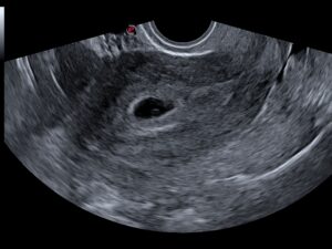
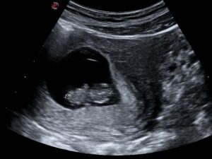
    At 4-5 weeks after the 1st day of the last menstrual period, we can only see a small gestation sac. (The fetus will not be visible as yet)

    At approximately 5 -6 weeks, we start to see the yolk sac in the gestation sac.

    7 Weeks

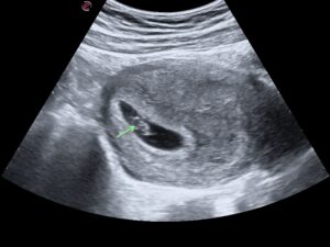
    At Approximately 7 weeks after the 1st day of the last menstrual period we will see a small fetus with a heartbeat in the gestation sac.

    10 Weeks

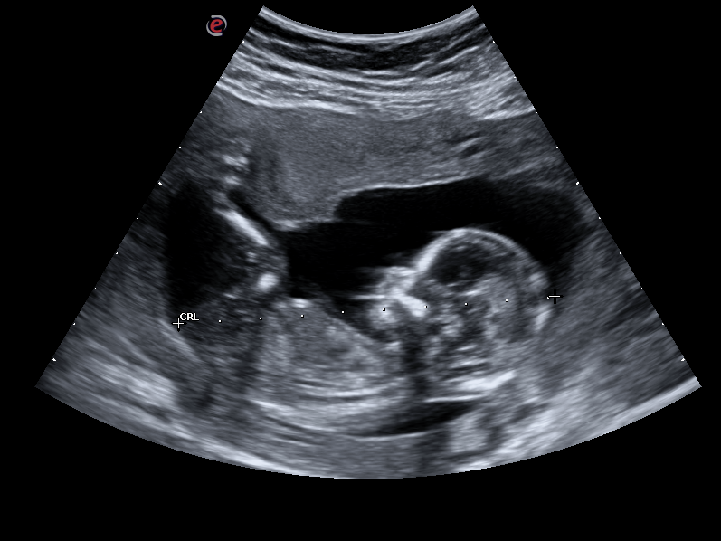
    At approximately 10 weeks we can start to see small arm and leg “buds”. Your baby will start to look like a jelly baby.

    Dating Ultrasound

    Most accurate time to determine the gestational age by ultrasound or how far you are in your pregnancy.

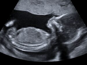
    A Nuchal translucency (fluid filled space at the back of the foetus´s neck) measurement can also be done at this time.

    A nuchal translucency measurement combined with other features of your baby in conjunction with a blood test (performed by Pathologist) can determine the risk for conditions such as Down´s syndrome.

    11 - 14 weeks

    Gender scan – From 16 weeks


    Boy
    Girl

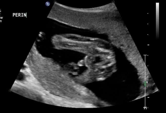
    The gender of the fetus can accurately be determed with ultrasound form 16 weeks gestation.

    Anomaly/Detailed or morphological Ultrasound

    (Please not this is not a 4D scan but a detailed 2D scan)
    Preferably done at 18-24 weeks, but can be done later if the gestation was missed.

    Heart
    Feet
    Hand

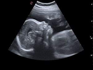
    A detailed ultrasound scan that looks at your baby’s body and organs but also observes the position of the placenta, the umbilical cord, the amniotic fluid.

    We will check to see if your baby has any abnormalities in their growth or development, including in their brain, face, heart, spine, stomach, kidneys bladder and limbs. Some of the abnormalities we aim to exclude are a cleft lip, spina bifida, a “hole” in the fetal heart, club feet ect.

    This type of scan is very important check that your baby is healthy. I value this scan the most important ultrasound in your pregnancy.

    Late / Third Trimester Ultrasound - 28 weeks to term.

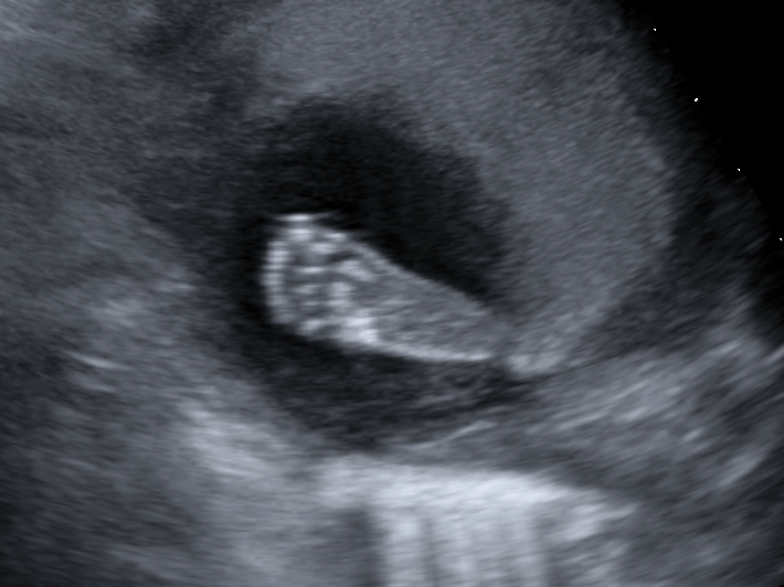
     These scans evaluate the fetal growth and to estimate the fetal weight. Confirm the position the fetus is lying. (If the head or the bum is down).  We will also verify the position of the placenta.  Doppler studies (ultrasound to determine blood flow) of the umbilical cord and/or the fetus brain can aslo be performed at this stage.

    Please note: “I am not a doctor” disclaimers:

  • An ultrasound is a diagnostic tool that may help with monitoring and/or making decisions regarding your pregnancy.  Your ultrasound will be performed by a qualified sonographer trained in obstetric ultrasounds.  I cannot and am not authorized to treat any condition.  Ultrasound is used to only monitor potential conditions and help with planning for further management.  If any problems or potential problems are detected you must seek medical attention for treatment at a healthcare provider of your choice.
  • Although the quality of most ultrasounds is excellent, and great care and time are put into every examination.  A number of disorders and defects cannot be identified with the use of ultrasound.  Factors that can limit the accuracy of ultrasound include but are not limited to, the age of your baby at the time of ultrasound, the positioning of the baby in the womb, your body type, and composition, and scarring left from previous surgeries.
  • From time to time it is not possible to image the necessary organs to make a complete evaluation and you might be rebooked (without further costs).
  • It is very important to know that some potential problems develop at different times in your pregnancy.  A normal ultrasound does not guarantee that a problem may not appear at a later date during your pregnancy.  A normal ultrasound also does not completely rule out potential birth defects, and does not guarantee that a genetic or chromosomal defect is absent.  Some birth defects cannot be detected with ultrasound.